Différences entre les versions de « Offres Traitement Images »
Aller à la navigation
Aller à la recherche
| Ligne 17 : | Ligne 17 : | ||
<tr> | <tr> | ||
<td> [[File:DTI.png|frameless|thumb| center|upright=1.2]]</td> | <td> [[File:DTI.png|frameless|thumb| center|upright=1.2]]</td> | ||
| + | <td> </td> | ||
<td> | <td> | ||
'''Estimation de cartes paramétriques '''<br> | '''Estimation de cartes paramétriques '''<br> | ||
| Ligne 24 : | Ligne 25 : | ||
<tr> | <tr> | ||
<td> [[File:corticalParcellation.png|frameless|thumb| center|upright=1.2]]</td> | <td> [[File:corticalParcellation.png|frameless|thumb| center|upright=1.2]]</td> | ||
| + | <td> </td> | ||
<td> '''Segmentation de structures anatomiques cérébrales''' | <td> '''Segmentation de structures anatomiques cérébrales''' | ||
<br> '''et de lésions de sclérose en plaques'''</td> | <br> '''et de lésions de sclérose en plaques'''</td> | ||
| Ligne 30 : | Ligne 32 : | ||
<tr> | <tr> | ||
<td> [[File:Atrophy.png|frameless|thumb| center|upright=1.2]]</td> | <td> [[File:Atrophy.png|frameless|thumb| center|upright=1.2]]</td> | ||
| + | <td> </td> | ||
<td> '''Estimation de l'atrophie cérébrale à un temps donnée'''<br> | <td> '''Estimation de l'atrophie cérébrale à un temps donnée'''<br> | ||
'''ou son évolution au cours du temps''' </td> | '''ou son évolution au cours du temps''' </td> | ||
| Ligne 36 : | Ligne 39 : | ||
<tr> | <tr> | ||
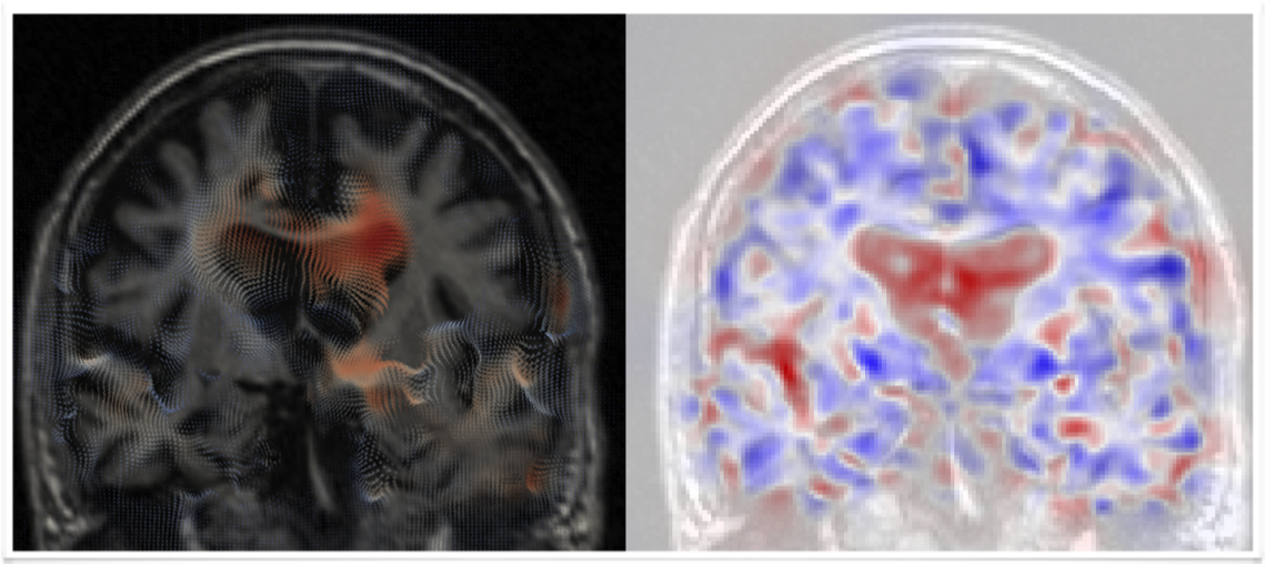
<td> [[File:ChangeDetection.png|frameless|thumb| center|upright=1.2]]</td> | <td> [[File:ChangeDetection.png|frameless|thumb| center|upright=1.2]]</td> | ||
| + | <td> </td> | ||
<td> '''Détection de changements entre deux examens'''<br> | <td> '''Détection de changements entre deux examens'''<br> | ||
''' d'IRM morphologique ou de diffusion''' </td> | ''' d'IRM morphologique ou de diffusion''' </td> | ||
| Ligne 42 : | Ligne 46 : | ||
<tr> | <tr> | ||
<td> [[File:fMRI.png|frameless|thumb| center|upright=1.2]]</td> | <td> [[File:fMRI.png|frameless|thumb| center|upright=1.2]]</td> | ||
| + | <td> </td> | ||
<td> '''Cartographie IRMf et l'extraction des réseaux de repos''' </td> | <td> '''Cartographie IRMf et l'extraction des réseaux de repos''' </td> | ||
<td> </td> | <td> </td> | ||
| Ligne 47 : | Ligne 52 : | ||
<tr> | <tr> | ||
<td> [[File:groupStudy.png|frameless|thumb| center|upright=1.2]]</td> | <td> [[File:groupStudy.png|frameless|thumb| center|upright=1.2]]</td> | ||
| + | <td> </td> | ||
<td>'''Comparaison de populations basée voxel ''' <br> | <td>'''Comparaison de populations basée voxel ''' <br> | ||
'''ou régions d'intérêt en IRM morphologique,'''<br> | '''ou régions d'intérêt en IRM morphologique,'''<br> | ||
Version du 9 décembre 2016 à 19:04
| Offres | Logiciels | Expertises |
En plus des moyens d'imagerie, la plate-forme offre une expertise et un service en traitement d'images médicales.
Le champ d'expertise couvre principalement l'imagerie cérébrale par IRM chez l'homme et le petit animal, mais peut être étendue à d'autres modalités d'imagerie (imagerie de médecine nucléaire, scanner X, ... ) et d'autres organes.
Catalogue de services
Les services proposés concernent notamment :
 |
Estimation de cartes paramétriques |
||
 |
Segmentation de structures anatomiques cérébrales
et de lésions de sclérose en plaques |
||
 |
Estimation de l'atrophie cérébrale à un temps donnée ou son évolution au cours du temps |
||
 |
Détection de changements entre deux examens d'IRM morphologique ou de diffusion |
||
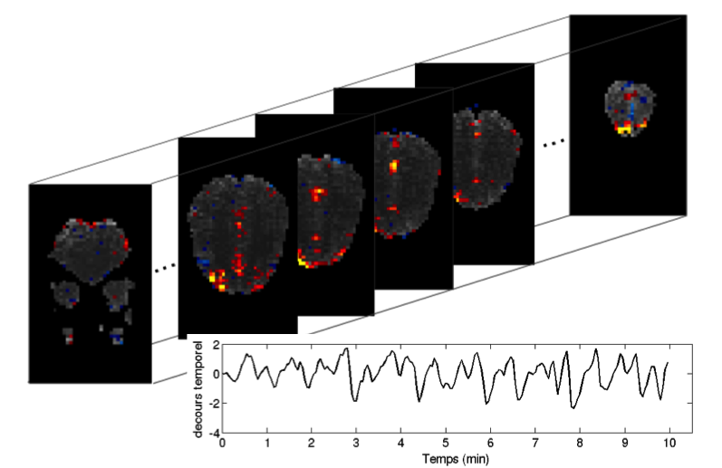 |
Cartographie IRMf et l'extraction des réseaux de repos | ||
 |
Comparaison de populations basée voxel ou régions d'intérêt en IRM morphologique, |
Par ailleurs, la plate-forme peut aussi offrir une formation et un support pour l'utilisation de différents logiciels de traitement d'images médicales (Medipy, SPM, FSL, ImageJ, ITK-Snap...)
Exemples de projets réalisés
- Étude de la mémoire autobiographique chez les sujets atteints de la maladie d’Alzheimer
- N. Philippi et al, "Different temporal patterns of specific and general autobiographical memories across the lifespan in Alzheimer’s disease", Behavioural Neurology, 2015(963460), 2015
- N. Philippi et al, "Impaired emotional autobiographical memory associated with right amygdalar-hippocampal atrophy in Alzheimer’s disease patients", Frontiers in Aging Neuroscience, 7(21), 2015.
- N. Philippi et al, "MRI-based volumetry correlates of autobiographical memory in Alzheimer", PLoS ONE, 7(10):e46200, 2012.
- Étude des hallucinations chez les sujets atteints de démences à corps de Lewy et de maladie d'Alzheimer
- C. Heitz et al, "Neural correlates of visual hallucinations in dementia with Lewy bodies", Alzheimer's Research and Therapy, 7(1), 2015.
- F. Blanc et al, "Right Anterior Insula: Core Region of Hallucinations in Cognitive Neurodegenerative Diseases", PLoS ONE, 9(12), 2014.
- Étude de la neuromyélite optique de Devic
- F. Blanc et al, "White matter atrophy and cognitive dysfunctions in neuromyelitis optica", PLoS ONE, 7(4) :33878, 2012.
- J. Chanson et al, "White matter volume is decreased in the brain of patients with neuromyelitis optica", European Journal of Neurology, 20(2), 2013.
- Étude des troubles olfactifs chez les sujets atteints du syndrome de Bardet Biedl
- J. Braun et al, "Value of MRI Olfactory Bulb evaluation in the assessment of olfactory dysfunction in Bardet Biedl syndrome", Clinical Genetics, 2016.
- J. Braun et al, "Olfaction evaluation and correlation with brain atrophy in Bardet Biedl syndrome", Clinical Genetics, 86(6), 2014.