Différences entre les versions de « Offres IRM Homme »
(Page créée avec « == Imagerie morphologique == == Imagerie paramétrique == == Angiographie == == IRM fonctionnelle (IRMf) == == Imagerie du tenseur de diffusion == == Imagerie en tem... ») |
|||
| (51 versions intermédiaires par 2 utilisateurs non affichées) | |||
| Ligne 1 : | Ligne 1 : | ||
| + | {| class="wikitable" style="font-size: 180%;" | ||
| + | |- | ||
| + | | [[Offres_IRM Homme|Offres]] || [[Équipement_IRM Homme|Équipements]] || [[Expertises_IRM Homme|Expertises]] | ||
| + | |} | ||
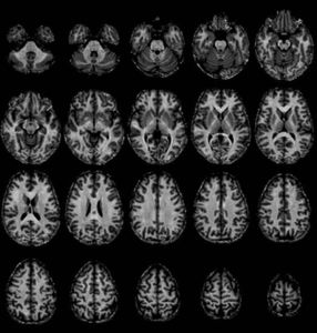
== Imagerie morphologique == | == Imagerie morphologique == | ||
| + | <gallery mode="packed-hover" heights="180"> | ||
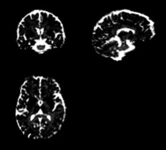
| + | File:3DT1_1mm.jpg|''T1w MPRAGE 1 mm isotropique'' | ||
| + | File:T1wMPR1iso0.7mm.jpg|''T1w MPRAGE 0,7 mm isotropique'' | ||
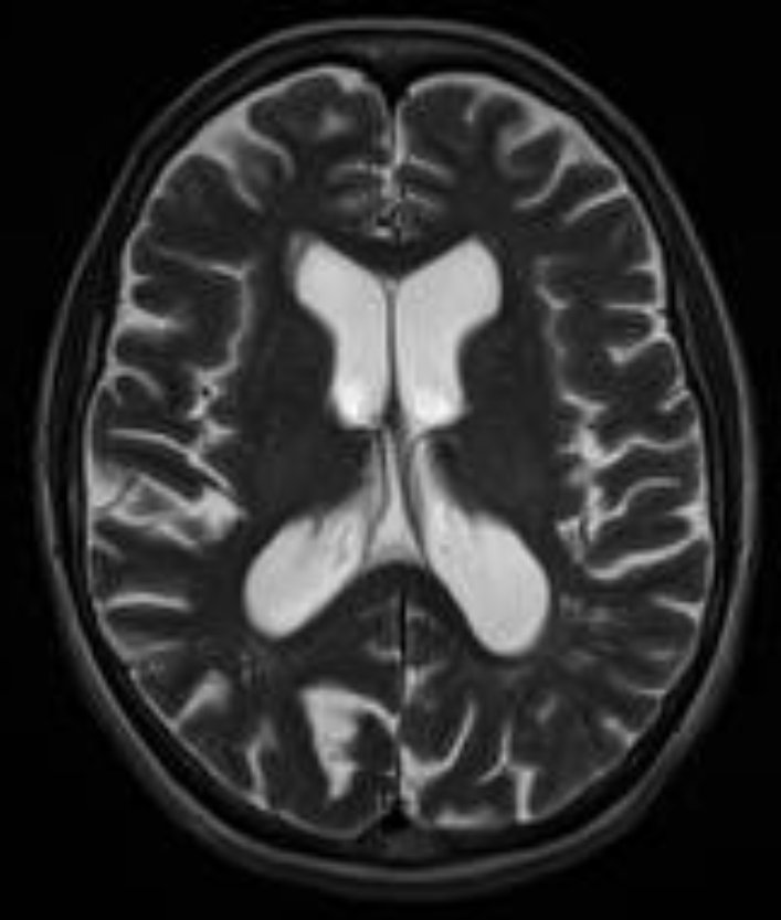
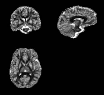
| + | File:T2TSE.jpg|''T2w TSE'' | ||
| + | File:T2GRE.jpg|''T2*w GRE'' | ||
| + | </gallery> | ||
| + | |||
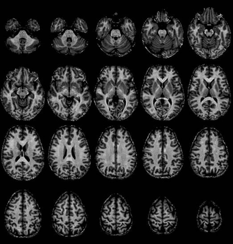
| + | <gallery mode="packed-hover" heights="180"> | ||
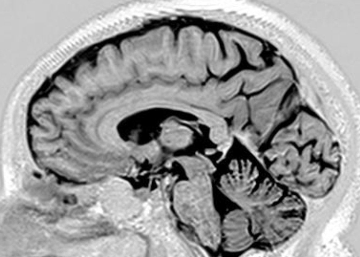
| + | File:T1_IR_SPC_SAG_ISO1mm_2.jpg|''T1w SPACE 1 mm isotropique'' | ||
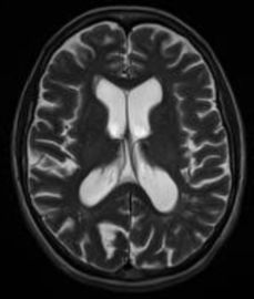
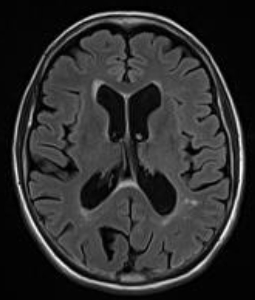
| + | File:T2FLAIR.jpg|''T2w FLAIR'' | ||
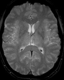
| + | File:mIP_SWI.jpg|''mIP SWI'' | ||
| + | </gallery> | ||
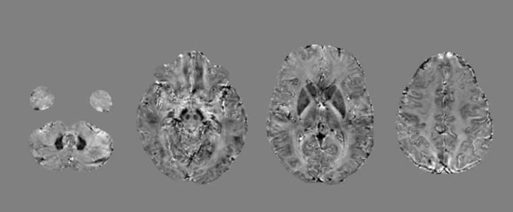
== Imagerie paramétrique == | == Imagerie paramétrique == | ||
| + | <gallery mode="packed-hover" heights="260"> | ||
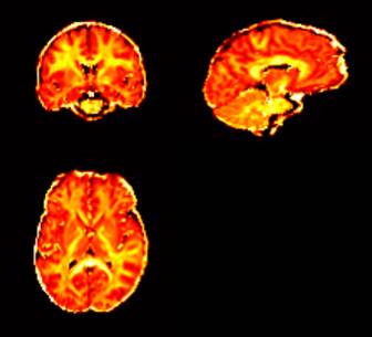
| + | File:T1map.jpg|''T1 map'' | ||
| + | File:T2map.jpg|''T2 map'' | ||
| + | File:T2star_map.jpg|''T2* map'' | ||
| + | </gallery> | ||
| + | |||
| + | <gallery mode="packed-hover" heights="200"> | ||
| + | File:qMTI_fmap.jpg|''Imagerie quantitative du Transfert d’aimantation'' | ||
| + | File:fast_QSM.jpg|''[[Imagerie de susceptibilité magnétique (QSM)]]'',- 4 minutes d'acquisition, 3D MGE, TR/TE1/TE8 = 37/2.21/28.11 ms, FA = 20°, 1 x 1 x 1 mm3 | ||
| + | </gallery> | ||
| + | |||
| + | == Imagerie vasculaire == | ||
| + | <gallery mode="packed-hover" heights="220"> | ||
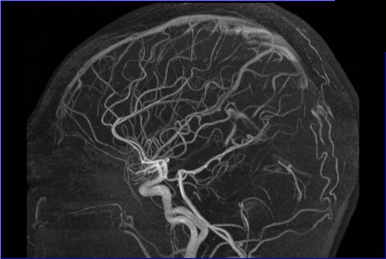
| + | File:TOF1.jpg|''Angiographie par temps de vol (TOF)'' | ||
| + | </gallery> | ||
| − | == | + | == Imagerie fonctionnelle (IRMf) == |
| + | <gallery mode="packed-hover" heights="240"> | ||
| + | File:ASL_BOLD_TR3s.jpg|''ASL BOLD TR 3 s'' | ||
| + | File:ASL_MULTIBAND.jpg|''BOLD Multi-Band TR 400 ms '' | ||
| + | </gallery> | ||
| − | == | + | == Imagerie de diffusion == |
| − | == | + | === Séquence Resolve (SIEMENS) === |
| + | <gallery mode="packed-hover" heights="250"> | ||
| + | File:RESOLVE_FA_color.jpg|''fraction d'anisotropie (FA)'' | ||
| + | </gallery> | ||
| + | |||
| + | <div style="text-align: center;"> | ||
| + | Séquence RESOLVE - 20 minutes d'acquisition : | ||
| + | TR/TE1/TE2 = 9400/83/108 ms, | ||
| + | 2 x 2 x 2 mm3, | ||
| + | 1b0 + 20 b1500 s/mm2 | ||
| + | </div> | ||
| + | |||
| + | === Neurite Orientation Dispersion and Density Imaging (NODDI) === | ||
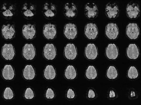
| + | <gallery mode="packed-hover" heights="200"> | ||
| + | File:NODDI1.jpg|''orientation dispersion index'' | ||
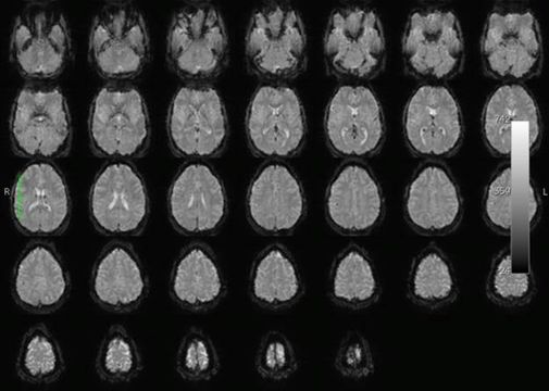
| + | File:NODDI2.jpg|''isotropic (CSF) volume fraction'' | ||
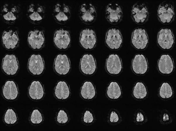
| + | File:NODDI3.jpg|''intra-cellular volume fraction'' | ||
| + | </gallery> | ||
| + | |||
| + | Protocole NODDI - 7 minutes d'acquisition : | ||
| + | 2D EPI SE-DIFF Multiband, TR/TE = 3460/94 ms, | ||
| + | FA = 78°, 2 x 2 x 2 mm3 | ||
| + | Multi-band accel. Factor = 3 | ||
| + | 12 b0 + 32 b700 + 64 b2000 s/mm2 | ||
== Imagerie en temps réel == | == Imagerie en temps réel == | ||
| + | |||
| + | [[Image:Real time.jpg|200px|link=https://youtu.be/3jc31YtEibo|Real time MRI]] | ||
| + | <-- cliquez ici | ||
Version actuelle datée du 4 janvier 2016 à 16:45
| Offres | Équipements | Expertises |
Imagerie morphologique
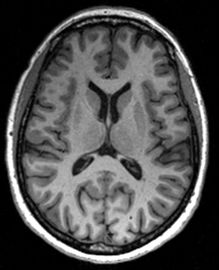
T1w MPRAGE 1 mm isotropique

T1w MPRAGE 0,7 mm isotropique
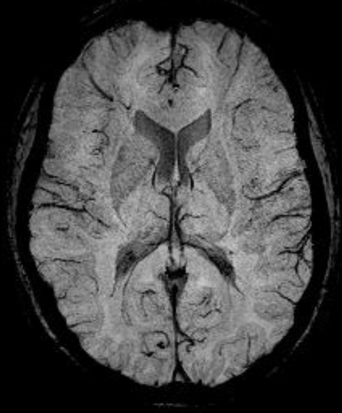
T2w TSE
T2*w GRE

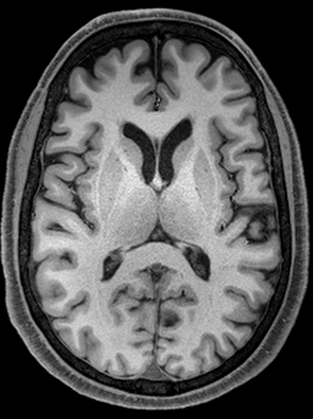


T1w SPACE 1 mm isotropique
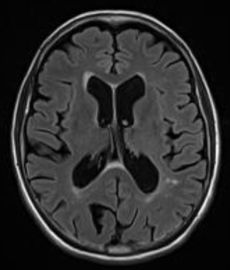
T2w FLAIR

mIP SWI
Imagerie paramétrique

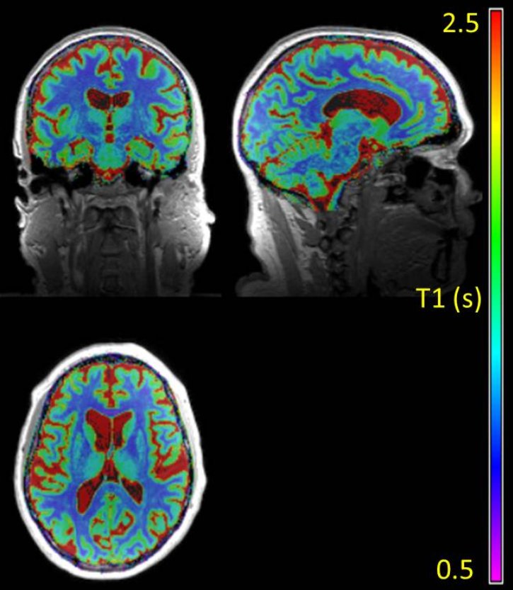
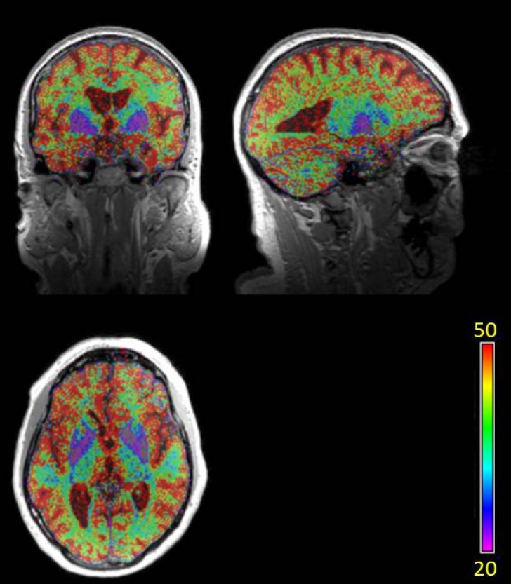
T1 map
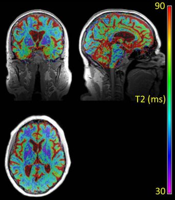
T2 map

T2* map

Imagerie quantitative du Transfert d’aimantation
Imagerie de susceptibilité magnétique (QSM),- 4 minutes d'acquisition, 3D MGE, TR/TE1/TE8 = 37/2.21/28.11 ms, FA = 20°, 1 x 1 x 1 mm3

Imagerie vasculaire
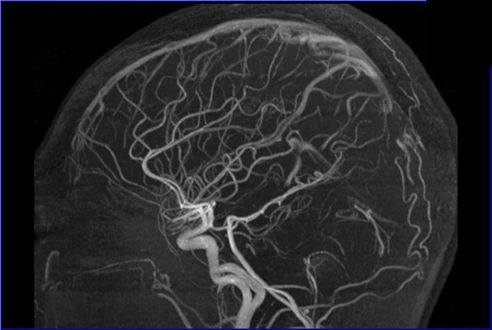
Angiographie par temps de vol (TOF)
Imagerie fonctionnelle (IRMf)
ASL BOLD TR 3 s
BOLD Multi-Band TR 400 ms

Imagerie de diffusion
Séquence Resolve (SIEMENS)
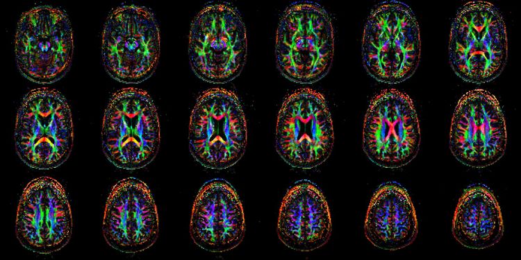
fraction d'anisotropie (FA)

Séquence RESOLVE - 20 minutes d'acquisition : TR/TE1/TE2 = 9400/83/108 ms, 2 x 2 x 2 mm3, 1b0 + 20 b1500 s/mm2
Neurite Orientation Dispersion and Density Imaging (NODDI)

orientation dispersion index
isotropic (CSF) volume fraction

intra-cellular volume fraction

Protocole NODDI - 7 minutes d'acquisition : 2D EPI SE-DIFF Multiband, TR/TE = 3460/94 ms, FA = 78°, 2 x 2 x 2 mm3 Multi-band accel. Factor = 3 12 b0 + 32 b700 + 64 b2000 s/mm2
Imagerie en temps réel
 <-- cliquez ici